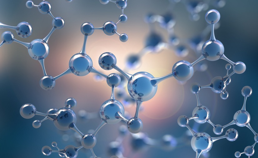
Figure 1: Abstract Molecular Model
Source: ShutterStock
Researchers at Nanyang Technological University (NTU) in Singapore have found a way to trick cancer cells into self-destructing, bypassing the need for radiation treatments, chemotherapy, or surgery. Their weapon? A nanoparticle (NP) secret agent that is 30,000 times smaller than a human hair.
These tiny particles come from the fast-evolving field of nanotechnology, the study of materials at an atomic scale in the range of zero to a hundred nanometers. A nanometer is a billionth of a meter. To put that into perspective, a piece of paper is 100,000 nanometers thick. At this size, physics behaves in strange ways, different from how we experience it in the macroscale. Capitalizing on the unique properties nano-materials exhibit has led to innovations in a variety of fields, especially in medicine.
Much of nano-oncology research has focused on using NPs as drug carriers, relying on the NP’s small size to reach tissues inaccessible through other methods. However, this approach is rife with complications—the tumors’ hostile environments can lower the drugs’ efficacy and accessibility, ultimately leading to increased incidences of drug resistance (Wu et al., 2020).
The NTU researchers side-stepped these issues, however, making their NPs cancer-killing weapons. They discovered that a silica nanoparticle cloaked in the amino acid phenylalanine—termed Nano-pRAAM, or Nanoscopic phenylalanine Porous Amino Acid Mimic—has cancer cell-destroying properties. Once inside a cancer cell, Nano-pRAAM over-activates the production of ROS (reactive oxygen species) molecules. While all cells have ROS molecules, cancer cells have higher ROS stress due to being more metabolically active than healthy cells (Pelicano et al., 2004). When the already intense ROS production in a cancer cell is stimulated further by the NP, it causes the cell to self-destruct. Dalton Tay, a material scientist at NTU Singapore, commented that, “’Against conventional wisdom, our approach involved using the nanomaterial as a drug instead [of] as a drug-carrier… Here, the cancer-selective and killing properties of Nano-pRAAM are intrinsic and do not need to be activated by any external stimuli’” (Nield, 2020).
The silica NP alone, though, cannot access the cancer cells; these malignant cells have to be tricked into absorbing the nanotherapeutic. Since cancer cells’ hyperactive metabolisms require a steady supply of exogenous amino acids, the NTA research team decided to cover their cancer-killing silica NP in phenylalanine. Not only does this act as a disguise fool the cancer cells into absorbing the NPs, this approach also selectively targets the cancer cells because they over-express a type of protein machinery—SLC7A5—that mediates the uptake of large amino acids. Thus, the nanotherapeutic leaves healthy tissue entirely alone (Wu et al., 2020).
Tested in the lab and on mice, this drugless approach killed 80% of gastric (stomach), breast, and skin cancer cells, a rate directly comparable with most modern treatments. Even better, the nanotherapeutic produced no noticeable side effects. Now, the researchers are looking to combine their approach with immunotherapy to reach even better outcomes. Hopefully, this kind of “Trojan horse sneak attack” may become a viable method for treating one of the most pernicious diseases known to humankind (Nield, 2020).
References
Nield, D. (2020, September 28). Experimental Cancer Treatment Destroys Cancer Cells Without Using Any Drugs. https://www.sciencealert.com/a-new-cancer-treatment-uses-a-trojan-horse-approach-and-no-drugs.
Pelicano, H., Carney , D., & Huang, P. (2004, April). ROS Stress in Cancer Cells and Therapeutic Implications. Science Direct. Retrieved from: https://www.sciencedirect.com/science/article/pii/S136876460400007X?via%3Dihub.
Wu, Zhuoran, Hong Kit Lim, Shao Jie Tan, Archana Gautam, Han Wei Hou, Kee Woei Ng, Nguan Soon Tan, and Chor Yong Tay. (August 2020). Potent‐By‐Design: Amino Acids Mimicking Porous Nanotherapeutics with Intrinsic Anticancer Targeting Properties. Small 16, no. 34: 2003757. https://doi.org/10.1002/smll.202003757.
Related Posts
Targeting crucial molecules used by coronaviruses to infect cells may lead to a broader antiviral treatment
Figure 1: Researchers have identified crucial molecules utilized by the...
Read MoreThe Mediterranean Diet Could Help Treat and Prevent Adolescent Depression
Source: Mediterranean Diet Pyramid (Flikr Public Domain, Zaid Alasad) The...
Read MorePANDAS––A Self-Destruction of the Mind
Figure 1: PANDAS, characterized by acute neurological and neuropsychiatric symptoms,...
Read MoreMary-Margaret Cummings


