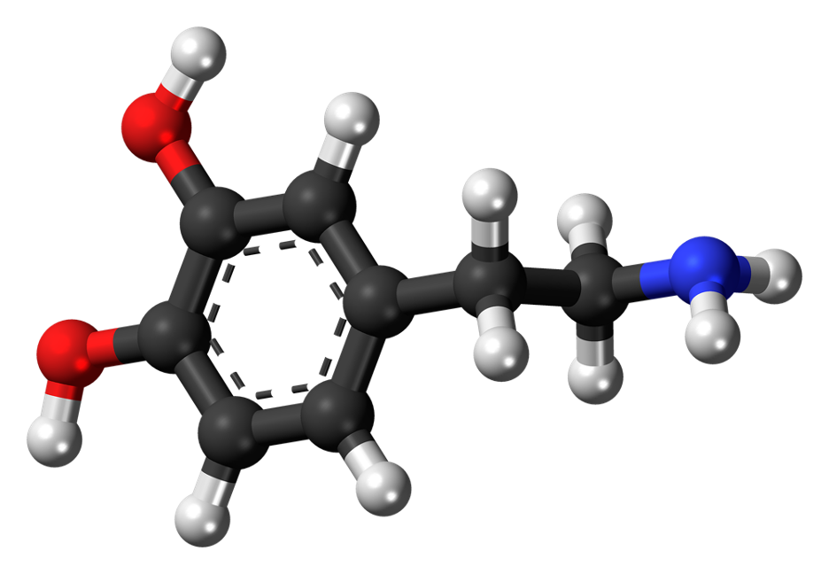
Figure: Dopamine is the so-called “feel-good” neurotransmitter, involved in cognitive processing including learning and behavioral planning (Foo et al., 2021). (Source: Jynto, Wikimedia Commons).
Traditionally, neural signaling has been studied in the context of “deterministic” signals, or external stimuli (Aguilera, 2021). However, a recent study by University of California San Diego researchers demonstrated that impulses of dopamine, the “feel-good” neurotransmitter, can also occur spontaneously (Aguilera, 2021; Foo et al., 2021). Furthermore, the study showed that mice were able to anticipate and willfully modulate these stochastic impulses, suggesting an internal awareness and control of extrasynaptic dopamine ([DA]ex) levels (Foo et al., 2021).
In this study, the researchers monitored extrasynaptic dopamine levels in the primary somatosensory cortex (where both deterministic and spontaneous dopaminergic signaling occur) using fluorescence resonant energy transfer (FRET), a type of fluorescent imaging used to show the localization of a tagged molecule (Foo et al., 2021; Sekar & Periasamy, 2003). Specifically, implanted cell-based neurotransmitter fluorescent-engineered reporters (CNiFERs) were used to produce an optical signal observed using in vivo two-photon microscopy (Foo et al., 2021).
The researchers first distinguished spontaneous dopamine impulses in the absence of a reward, observing a wide distribution of both impulse length and amplitude (Foo et al., 2021). Then, they measured spontaneous dopamine impulses at baseline (no reward), given feedback (sucrose-water given in response to increased [DA]ex), then without feedback again (no reward). Within the second stage, referred to as the “feedback ON” stage, the researchers observed changes in [DA]ex both when the mice were rewarded simply for [DA]ex above a determined baseline and when the reward threshold either increased or decreased in accordance with [DA]ex change (Foo et al., 2021).
By the second day of feedback training, the researchers observed a statistically significant increase in [DA]ex (Foo et al., 2021). When the feedback was removed on day 4, the [DA]ex level returned to baseline, but it was restored with a return of feedback on day 5, demonstrating that the mice’s learned ability to volitionally control [DA]ex was not extinguished by one day without reward. The same effect that occurred on day 4 occurred again on day 6 when feedback was once again omitted (Foo et al., 2021).
The increased amplitude and duration of spontaneous dopamine impulses in the mice provided with neurofeedback in the form of a reward suggests that the mice were able to learn to volitionally control [DA]ex independently of sensory stimuli (Foo et al., 2021). This finding could be significant for advances in neuromodulation therapy in humans, which is the control of neural activity using targeted stimulus delivery (International Neuromodulation Society, 2014). Neuromodulation therapy is used to treat brain disorders such as chronic neuropathic pain, Parkinson’s disease, and epilepsy, or mental illnesses such as depression and obsessive-compulsive disorder (International Neuromodulation Society, 2014). In major depressive disorder (MDD) in particular, the dysregulation of afferent dopaminergic neurons (those releasing extrasynaptic dopamine) is thought to be one of the main pathophysiological mechanisms (Belujon & Grace, 2017). Several studies have investigated the use of neurofeedback for treatment of MDD, specifically monitoring brain wave patterns (using electroencephalogram, or EEG) or dopaminergic signaling (using real-time functional magnetic resonance imaging, or rtfMRI) (Lee et al., 2019; Cheon et al., 2016; Trambaiolli et al., 2021; Sulzer et al., 2013). Foo and colleagues’ finding that spontaneous dopamine impulses can be volitionally controlled using neurofeedback may further reinforce the promise of neuromodulation therapies for MDD and other brain disorders featuring dopaminergic dysregulation (2021).
References
- Aguilera, Mario. (2021, July 23). “Feel good” brain messenger can be willfully controlled, new study reveals: Neuroscientists show that mice can learn to manipulate random dopamine impulses for reward. ScienceDaily. https://www.sciencedaily.com/releases/2021/07/210723121512.htm
- Belujon, P., & Grace, A. A. (2017). Dopamine System Dysregulation in Major Depressive Disorders. International Journal of Neuropsychopharmacology, 20(12), 1036–1046. https://doi.org/10.1093/ijnp/pyx056
- Cheon, E.-J., Koo, B.-H., & Choi, J.-H. (2016). The Efficacy of Neurofeedback in Patients with Major Depressive Disorder: An Open Labeled Prospective Study. Applied Psychophysiology and Biofeedback, 41(1), 103–110. https://doi.org/10.1007/s10484-015-9315-8
- Foo, C., Lozada, A., Aljadeff, J., Li, Y., Wang, J. W., Slesinger, P. A., & Kleinfeld, D. (2021). Reinforcement learning links spontaneous cortical dopamine impulses to reward. Current Biology, 0(0). https://doi.org/10.1016/j.cub.2021.06.069
- International Neuromodulation Society. (2013). Neuromodulation, or Neuromodulatory Effect. International Neuromodulation Society. https://www.neuromodulation.com/neuromodulation-defined
- Lee, Y.-J., Lee, G.-W., Seo, W.-S., Koo, B.-H., Kim, H.-G., & Cheon, E.-J. (2019). Neurofeedback Treatment on Depressive Symptoms and Functional Recovery in Treatment-Resistant Patients with Major Depressive Disorder: An Open-Label Pilot Study. Journal of Korean Medical Science, 34(42), e287. https://doi.org/10.3346/jkms.2019.34.e287
- Sekar, R. B., & Periasamy, A. (2003). Fluorescence resonance energy transfer (FRET) microscopy imaging of live cell protein localizations. The Journal of Cell Biology, 160(5), 629–633. https://doi.org/10.1083/jcb.200210140
- Sulzer, J., Sitaram, R., Blefari, M. L., Kollias, S., Birbaumer, N., Stephan, K. E., Luft, A., & Gassert, R. (2013). Neurofeedback-mediated self-regulation of the dopaminergic midbrain. NeuroImage, 83, 817–825. https://doi.org/10.1016/j.neuroimage.2013.05.115
- Trambaiolli, L. R., Kohl, S. H., Linden, D. E. J., & Mehler, D. M. A. (2021). Neurofeedback training in major depressive disorder: A systematic review of clinical efficacy, study quality and reporting practices. Neuroscience and Biobehavioral Reviews, 125, 33–56. https://doi.org/10.1016/j.neubiorev.2021.02.015
Related Posts
Human Endogenous Retroviruses Might Unlock a New Field of Neurodegenerative Disease Research
Figure 1: Researchers have found viral genetic materials within the...
Read MoreStrength in Numbers: How Community Enhances Perceived Support
Figure 1: Bandmates join in a group hug. The powerful...
Read MoreCellChat Enables Biologists to Understand Cellular Communication
Figure 1: CellChat, an open-source R package, provides researchers with...
Read MoreNew Study Finds that Media Multitasking Impairs Memory in Young Adults
Figure 1: Engaging with multiple types of digital media simultaneously,...
Read MoreA New Method to Track Parkinson’s Disease
Figure 1: 3D structure of alpha-synuclein protein. Mutations in alpha-synuclein...
Read MoreUniversality of the Face: New Research Points to a Shared Human Commonality in Emotional Expression
Figure 1: Examples of emotional expressions Source: Andrea Piacquadio from Pexels Raising...
Read MoreJillian Troth


