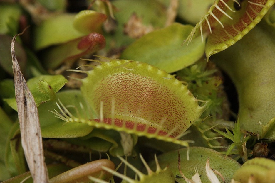
Figure 1: An image of a Venus Flytrap plant (Dionaea muscipula)
Source: Wikimedia Commons
A multidisciplinary team of researchers have been able to measure magnetic fields produced by a multicellular plant for the first time without the use of invasive or supercooled magnetometers. This finding is important despite the fact that magnetic fields have been detected in single-cell “plants” (like protists) and multicellular animals (this is the basis for many medical diagnostic tools) because the field of plant magneto-physiology is relatively new (ScienceDaily, 2021).
In 1820, Hans Christian Oersted demonstrated that the origin of magnetic fields is the flow of charges, or current. Oersted observed that a wire carrying charge was able to move the needle of his compass. Over time, our understanding of electric and magnetic fields, their accompanying forces, and the electromagnetic radiation (aka, light) caused by disturbances in these fields has become far more sophisticated. In 1963, Baule and McAfee were able to prove that the human heart generates a magnetic field due to the electrical impulses that keep it beating. From there, the concept of biological action potentials, the flow of charged ions between the interior and exterior of cells (like neurons, muscle cells, and hair cells on Venus Flytraps) that are used for stimulus transduction, led many to believe that this form of biological current should generate magnetic fields. This idea formed the basis of the many contemporary diagnostic tools such as electroencephalography (EEG), magnetoencephalography (MEG), and magnetic resonance imaging (MRI), which rely on magnetic and electrical impulses generated in the human body (Williamson & Kaufman, 1981).
Scientists have previously detected magnetic fields generated specifically in plants (from electrical signals they use for stimuli transduction, similar to animals), but only with equipment that required either the placement of electrodes directly into the plant’s cells or the complicated process of supercooling, such as experiments conducted with the superconducting-quantum-interference-device (SQUID). Due to the usefulness of the non-invasive, cryogenically-cooled diagnostic tools that detect biomagnetic fields in humans using massive magnets, making analogous tools for plants has been a prominent goal of the agricultural industry. However, plant damage, and the costly process of cryogenic cooling (the cost of which is less troublesome when it comes to using the technology to diagnose and help people) with the SQUID device has largely prevented such tools from being developed. To combat these problems, recent researchers measured the biomagnetic fields of plants using optically pumped atomic magnetometers, which employ lasers to alter the spin state of electrons of vaporized alkali metals (i.e., to “optically pump” them), thus creating a gaseous material within the plant that is highly magnetically sensitive (Tierney et al., 2019).
Past research has determined that the action potential which propels the closing of the Venus Flytrap’s trap can be stimulated via heat. The heat provides energy to stimulate the opening of calcium (Ca2+) channels in the hair cells of the plant. This mechanism causes a cellular influx of Ca2+, allowing for activation of the Ca2+ dependent anion channels on the cell membrane and a subsequent efflux of anions, generating an action potential. To minimize worry about the possible confounding effects of stimulating the hair cells manually or with actual prey, the research team led by Anne Fabricant used isolated traps and determined the proper temperature (greater than 40℃) to cause trap closures and thus generating action potentials (2021). They then set these isolated traps in magnetically shielded rooms, stimulated action potentials via heat, and measured the magnetic field generated by these action potentials via optically pumped atomic magnetometers at different angles and spacing around the traps (Fabricant et al., 2021).
In doing so, the research team determined the strength of the magnetic field generated by the action potential to be around 0.5pT (a fact that they checked by using the estimated current caused by an action potential to calculate the estimated field strength, which was about 0.3pT). Additionally, this number, while millions of times smaller than the magnetic field strength of the Earth (generated by the current produced by the flow of motel ions in the Earth’s core), is similar to signals detected within humans and other animals (Fabricant et al., 2021).
While researchers had to take these measurements in a magnetically shielded room, this technique has the potential be used in developing a database of specific magnetic fields generated by a given plant in response to stimulus. This advance in technology would allow farmers to determine optimal temperatures for their crops, discover parasitic crop infections, and even verify if a pesticide is toxic to a crop field.
References
Fabricant, A., Iwata, G. Z., Scherzer, S., Bougas, L., Rolfs, K., Jodko-Władzińska, A., Voigt, J., Hedrich, R., & Budker, D. (2021). Action potentials induce biomagnetic fields in carnivorous Venus flytrap plants. Scientific Reports, 11(1), 1438. https://doi.org/10.1038/s41598-021-81114-w
Johannes Gutenberg Universitaet Mainz. (2021, February 2). Venus flytraps found to produce magnetic fields: Physicists use atomic magnetometers to measure the biomagnetic signals of the carnivorous plant. ScienceDaily. Retrieved February 23, 2021 from www.sciencedaily.com/releases/2021/02/210202113815.htm
Tierney, T. M., Holmes, N., Mellor, S., López, J. D., Roberts, G., Hill, R. M., Boto, E., Leggett, J., Shah, V., Brookes, M. J., Bowtell, R., & Barnes, G. R. (2019). Optically pumped magnetometers: From quantum origins to multi-channel magnetoencephalography. NeuroImage, 199, 598–608. https://doi.org/10.1016/j.neuroimage.2019.05.063
Williamson, S. J., & Kaufman, L. (1981). Biomagnetism. Journal of Magnetism and Magnetic Materials, 22(2), 129–201. https://doi.org/10.1016/0304-8853(81)90078-0
Related Posts
Insect Microbiome Gives Hope for New Antibiotic Discovery
Figure 1: Actinobacteria were isolated from 56% of the insect...
Read MoreBio-renewable Alternative to Polyethylene Terephthalate (PET)
The pliability and low-cost of synthetic thermoplastics have rendered them...
Read MoreNew Wearable, Body Heat Fueled Generator With Self-Repairing Capabilities
Figure 1: This is a photograph of several current wearable...
Read MoreFrankie Carr


