Cover Image: The Structure of the Coronavirus. (Source: Wikimedia Commons, Khera)
Introduction
Originating in 2019 in a province called Wuhan, China, the Coronavirus Disease (COVID-19) has spread like wildfire, leaving behind a trail of economic downturn, health crises and poverty. According to the World Health Organization, this novel disease has infected over ten million people and has caused over 4 million deaths worldwide (World Health Organization, 2021). The rapid spread of COVID-19 can be owed to its communicable nature. It is severely contagious and can be transmitted from person to person by means of droplets, aerosols, and surfaces (Jayaweera et al., 2020). This transmission occurs because particles of the virus are condensed into globs of mucus, saliva, and water and are released into the environment whenever an infected person talks, coughs or sneezes (Jayaweera et al., 2020). Any individual within the vicinity of an infected person will thereafter inhale the released particles and contract the disease.
COVID-19 typically settles in the lungs, making it a respiratory disease. After inhalation, the virus – assisted by the spike glycoprotein located on its outer surface – enters the epithelial cells of the lungs, and hinders their function (Sadhukhan et al., 2020). Thus, the most common symptoms observed are breathlessness, chest pain, cough, and nasal congestion. Although COVID-19 affects the lungs most disastrously, it has been found to impact other organs as well. A journal in the Netherlands – The Journal of Pathology – reports that COVID-19 can result in instances of myocardial (heart muscle) injury caused by elevated levels of cardiac troponin T in the body. Furthermore, the same journal reports gastrointestinal, renal (kidney), as well as hepatic (liver) effects of COVID-19 in patients as well (Bourgonje at al., 2020). The gastrointestinal symptoms are caused directly by the virus and typically manifest in the form of abdominal pain, diarrhea, and nausea, while renal and hepatic complications arise mostly due to the high dosages of medications and antibiotics administered as a treatment for this disease. Finally, the central topic of this article. COVID-19 affects the brain in many ways. Reduced supply of oxygen to the brain can cause brain fog or mental confusion and can even result in stroke. Moreover, memory deficits linked to the anosmia caused by the disease and epileptic seizures are also commonly seen.

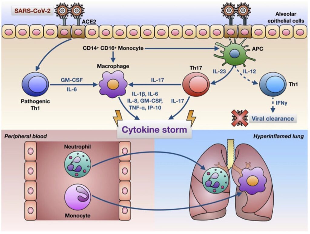
Figure 1: The Mechanisms Involved in the Cytokine Storm Triggered by COVID-19. (Source: Wikimedia Commons, Corrado)
Attention – Deficits caused by COVID-19
Multiple studies have uncovered that COVID-19 causes several unusual symptoms in patients. One such symptom is known as ‘brain fog,’ or dysexecutive syndrome (Nauen, 2021). Brain fog is characterized by deficits in short term memory, confusion, headaches, and an inability to focus or pay attention to one’s surroundings (Memorial Sloan Kettering Cancer Center, 2021).
Researchers at the Memorial Sloan Kettering Cancer Center speculate that this brain fog is not directly caused by the virus but is caused by the presence of inflammatory molecules in the cerebrospinal fluid which surrounds the brain (Memorial Sloan Kettering Cancer Center, 2021). The Sloan-Kettering scientists examined samples of the cerebrospinal fluid of COVID-19 patients and concluded that the delirium could be due to the oft-mentioned cytokine storm. A cytokine storm is a phenomenon in which a virus or foreign body causes one’s immune system to go into overdrive, resulting in an overflow of immune proteins in the bloodstream. The result is often damage to tissues, which can thereby corrupt the different organs in the body, including the brain, leading to delirium (Pathak, 2021). Adding to this research, the department of medicine at Johns Hopkins University claims that bone marrow cells called megakaryocytes specifically cause this impaired awareness. The cytokine storm in the cerebrospinal fluid can trigger the migration of megakaryocytes to the brain, blocking the flow of fresh, oxygenated blood to the cerebral cortex producing brain fog. Furthermore, cytokines can directly cause inflammation in local areas, thus hindering the communication between neurons in the brain (White, 2021).
While the aforementioned findings offer a biological explanation of the cause of brain fog, a study conducted in China highlights the psychological contributors of brain fog in COVID-19 patients. According to this study, 20% of patients experienced insomnia, 18.5% developed anxiety and approximately 24.5% suffered from depression following COVID-19. All these conditions contributed to brain fog in patients (Morin & Carrier, 2021). Furthermore, this study claims that the disruption caused by COVID-19 in people’s daily routines, and the lack of natural sunlight – a potential consequence of the ‘stay at home’ mandate observed in most countries – has interfered with the proper functioning of the circadian rhythm, thereby contributing to insomnia (Morin & Carrier, 2021). Maintaining a proper routine regulates the circadian rhythm of the body. However, the social isolation and confinement experienced by COVID-19 patients disrupts this daily routine, throwing the sleep-wake cycle off, leading to insomnia. This insomnia can thereby snowball into anxiety and depression. Additionally, the financial problems, social seclusion, and the threat to one’s life and health that have come along with the pandemic contribute to COVID-19 related anxiety and depression.
Epileptic Seizures triggered by COVID-19
Epilepsy is a prevalent medical condition, affecting over 50 million individuals worldwide, annually (World Health Organization, 2020). This condition can be caused by severe neuronal excitability driven by the excess release of neurotransmitters like glutamate, or a decrease in the release of inhibitory neurotransmitters like gamma-aminobutyric acid (GABA). Epileptic seizures can also be caused by other dysfunctions such as head trauma, fevers, inflammation, hypoglycemia, or hyperglycemia (Mayo Foundation for Medical Education and Research, 2021). Recent research on the coronavirus pandemic suggests that COVID-19 can also be a trigger for epileptic seizures. It was found that approximately 1.2% of COVID-19 patients had an epileptic seizure post recovery (Vossler, 2021). The virus causes epileptic seizures by affecting the nervous system in four ways – triggering a cytokine storm, impairing the blood brain barrier, disrupting the functioning of the mitochondria, and by producing an electrolyte imbalance.
Not only does a cytokine storm cause brain fog and attention deficits, but it also may epileptic seizures. When the virus enters the body, it can reach the central nervous system by infecting the neurons which relay information to and from the brain and spinal cord. After the infection, the virus promotes a cytokine storm which causes inflammation in the area. This inflammation can upset the neurotransmitter balance in the brain by triggering the release of too much glutamate and too little GABA in the synapses between neurons, contributing to the onset of a seizure (Nikbakht et al., 2020).
COVID-19 has also been known to disrupt the proper functioning of the blood brain barrier (BBB), which can cause seizures as well (Nikbakht et al., 2020). When the virus permeates the BBB, it causes the movement of blood cells and proteins away from the region, upsetting the osmotic balance in the area. This imbalance can cause seizures in the brain (Nikbakht et al., 2020). COVID-19 also causes hyperthermia and fever in infected individuals which is known to adversely affect BBB integrity (Nikbakht et al., 2020). Thus, the coronavirus is capable of causing seizures by this more indirect mechanism also.
On a cellular level, the involvement of the mitochondria in the pathology of COVID-19 is two-fold. From generating energy, to producing reactive oxygen species (ROS), the mitochondria serve many functions which are necessary for regulating the body’s metabolism (Nikbakht et al., 2020). When the body is infected with the COVID-19 virus, the inflammatory cytokine storm causes an increase in the production of ROS by the mitochondria. This proves to be harmful because the growth of ROS above baseline rates can contribute to the pathogenesis of autoimmune disease and cardiomyopathies. Furthermore, the dysfunction of the mitochondria disables it from performing its chief function – energy production – which is necessary for proper electrical signaling in the brain. A fault in the conduction of impulses between neurons can also lead to epilepsy and seizures (Nikbakht et al., 2020).
Lastly, studies have deciphered a connection between electrolyte imbalances and COVID-19. The virus is known to disturb the functioning of the ACE 2 receptor, which leads to an increase in the concentration of angiotensin II in the body. The increased concentration of angiotensin II, in turn, forces the kidneys to excrete more electrolytes like potassium, magnesium, and calcium, – which are essential for the transmission of impulses in the brain (Nikbakht et al., 2020). This results in conditions like hypokalemia, hypomagnesemia, and hypocalcemia which can cause epileptic seizures and brain damage.
Memory Deficits, Anosmia, and COVID-19
One of the most reported symptoms of COVID-19 is anosmia or the loss of the sense of smell. While this sense is usually recovered as patients convalesce from the disease, studies report that anosmia can have lasting impacts on the brain. A study conducted in the United Kingdom reports that COVID-19 can cause a loss of gray matter in the cortical areas of the limbic system which are associated with the olfactory and the gustatory pathways of the body (Gwenaëlle et al., 2021).
The olfactory pathway begins in the nasal cavity of the nose, which is lined with olfactory receptors. From there, the nerve fibers of the pathway connect the receptors in the nasal cavity and carry input to the olfactory bulb in the forebrain. Recent research suggests that the cells in this system are sensitive to COVID-19, thus the olfactory mucosa and the olfactory bulb can become a point of entry of the virus into the central nervous system (Gwenaëlle et al., 2021). The bulb also connects to different regions of the limbic system such as the piriform cortex, the parahippocampal gyrus, the entorhinal cortex, and the amygdala – regions which are responsible for the formation and the consolidation of memories in the brain (Britannica, T. Editors of Encyclopaedia, 2019). Thus, as the virus effects the olfactory bulb, and consequently the sense of smell, it also affects the thickness and volume of the gray matter in the limbic system, thereby compromising the patient’s ability to consolidate memories (Gwenaëlle et al., 2021).
The memory loss caused by COVID-19 is also associated with the inflammatory response set off by the body’s immune system and its consequent neuroplastic effect (Bhardwaj, 2021). In response to a local infection, the immune system signals to the neuro-immune cells in the brain that they need to counteract the virus. This immune response has an inflammatory effect on the brain and can modify the way neurons send signals and communicate with each other, thus interfering with the storage of information. Moreover, certain neuro-immune cells can also settle in the synaptic clefts between neurons, hindering the formation of synapses and the encoding of new memories.
Researchers in Norway conducted a study to better understand the link between COVID-19 and memory loss (Søraas et al., 2021). For the study, patients who had tested positive for COVID-19 were recruited and asked about their symptoms, underlying medical conditions, and demographic information, and were made to answer a baseline questionnaire. Eight months after their recovery from the disease, patients were asked if they experienced difficulty remembering things and if they had experienced episodes of cognitive deficits by way of confusion or trouble concentrating. Out of the 75% of participants who responded to the follow up, about 11% reported problems remembering things, while 12% reported difficulty in concentrating. This implies that the novel coronavirus can have a lasting impact on the brain and cognition (Søraas et al., 2021).
Conclusion
Although COVID-19 is primarily a respiratory disease, a large body of research implicates the virus as an agent that can cause dysfunction of the brain and the nervous system. From attention deficits and memory problems to epileptic seizures, the virus can cause a plethora of unusual symptoms in patients which are still being understood. Inflammation triggered by a cytokine storm plays a notable role. The cytokine storm can cause the migration of megakaryocytes to the region of infection, blocking the flow of oxygen to the brain causing brain fog. It can also cause a neurotransmitter imbalance, resulting in epileptic seizures. Lastly, inflammation can hinder the synaptic connections between neurons, thus preventing the encoding of new memories. In addition, the virus infiltrates the olfactory system causing anosmia and memory loss, disturbs mitochondrial functioning, and even permeates the blood brain barrier causing seizures.
The virus can also have long lasting psychological effects on patients, as it triggers anxiety, depression, and stress. However, practices like mindfulness meditation have proven to be successful in combating these problems, enhancing mood, and facilitating memory retrieval (Vatansever et al., 2021). Regular meditation and mindfulness training can induce positive structural changes in the brain improving attention and cognition in those recovering from the disease. Furthermore, mindfulness meditation and stress reduction therapies have been linked with an increase in gray matter density in the brain as observed by the Harvard Medical School (Lewis, 2019). Therefore, despite all the negative effects COVID-19 has brought, there is still hope for a strong recovery in affected individuals.
References
Bhardwaj, M. (2021, February 12). Can COVID-19 really cause Memory LOSS?: AAKASH HEALTHCARE. Can COVID-19 Really Cause Memory Loss? | Aakash Healthcare. https://aakashhealthcare.com/can-covid-19-really-cause-memory-loss.
Bourgonje, A. R., Abdulle, A. E., Timens, W., Hillebrands, J. L., Navis, G. J., Gordijn, S. J., Bolling, M. C., Dijkstra, G., Voors, A. A., Osterhaus, A. D., van der Voort, P. H., Mulder, D. J., & van Goor, H. (2020). Angiotensin-converting enzyme 2 (ACE2), SARS-CoV-2 and the pathophysiology of coronavirus disease 2019 (COVID-19). The Journal of pathology, 251(3), 228–248. https://doi.org/10.1002/path.5471
Britannica, T. Editors of Encyclopaedia (2019, October 11). Olfactory system. Encyclopedia
Britannica. https://www.britannica.com/science/olfactory-system
Gwenaëlle D., Soojin L. , Fidel A.A. , Christoph A. , Chaoyue W., Frederik L., Jesper L.R. A., Ludovica G., Eugene., Saad J., Bernd T., Anderson W., Thomas E.N., Rory C., Paul M.M., Naomi A., Karla L. M., Stephen M. S. (2021). Brain Imaging Before and After COVID-19 in UK Biobank. MedRxiv.
Jayaweera, M., Perera, H., Gunawardana, B., & Manatunge, J. (2020). Transmission of COVID-19 virus by droplets and aerosols: A critical review on the unresolved dichotomy. Environmental research, 188, 109819. https://doi.org/10.1016/j.envres.2020.109819
Kausalya (2021). 3D Medical Animation Showing Still Shot Coronavirus [Infographic]. ScientifiAanimations.com. https://www.scientificanimations.com/3d-medical-animation-still-shot-showing-coronavirus-structure/
Koshy, J. (2021, June 24). Study finds loss in grey matter after coronavirus infection. The Hindu. https://www.thehindu.com/sci-tech/health/study-finds-loss-in-grey-matter-after-coronavirus-infection/article34936719.ece.
Lewis, B. (2019, June 13). Mindfulness increases gray matter density. mind body seven. https://www.mindbody7.com/news/2017/10/24/mindfulness-practices-lead-to-increased-gray-matter-density#:~:text=Studies%20suggest%20that%20mindfulness%20practices,referential%20processing%20and%20perspective%20taking.
Marshall, M., (2020). How COVID-19 can damage the brain. Nature 585, 342 – 343. doi: https://doi.org/10.1038/d41586-020-02599-5
Memorial Sloan Kettering Cancer Center (2021, February 8). MSK Researchers Learn What’s Driving ‘Brain Fog’ in People with COVID-19. https://www.mskcc.org/news/msk-researchers-learn-what-s-driving-brain-fog-people-covid-19.
Morin, M. C., Carrier, J., Bastien, C., Godbout, R. (2020). Sleep and circadian rhythm in response to the COVID-19 pandemic. Canadian Journal of Public Health, 111(5): 654-657. doi: 10.17269/s41997-020-00382-7
Morin, C. M., & Carrier, J. (2021). The acute effects of the COVID-19 pandemic on insomnia and psychological symptoms. Sleep medicine, 77, 346–347. https://doi.org/10.1016/j.sleep.2020.06.005
Mayo Foundation for Medical Education and Research. (2021, March 31). Epilepsy. Mayo Clinic. https://www.mayoclinic.org/diseases-conditions/epilepsy/symptoms-causes/syc-20350093.
Nauen, D. (2021, February 24). Covid-19 story Tip: Researchers may HAVE UNCLOUDED the mystery of COVID-19 ‘brain fog’. Johns Hopkins Medicine Newsroom. https://www.hopkinsmedicine.org/news/newsroom/news-releases/covid-19-story-tip-researchers-may-have-unclouded-the-mystery-of-covid-19-brain-fog.
Nikbakht, F., Mohammadkhanizadeh, A., & Mohammadi, E. (2020). How does the COVID-19 cause seizure and epilepsy in patients? The potential mechanisms. Multiple sclerosis and related disorders, 46, 102535. https://doi.org/10.1016/j.msard.2020.102535
Patel, R., Rinker, L., Peng, J., Chilian, M. W. (2017). Reactive Oxygen Species: The Good and the Bad. IntechOpen. http://dx.doi.org/10.5772/intechopen.71547
Pathak, N. (Ed.). (2021, August 23). Coronavirus and COVID-19: What you should know. WebMD. https://www.webmd.com/lung/coronavirus.
Pelaia C. (2021). Hypothetical Mechanisms Underlying the Cytokine Storm Induced by SARS-CoV -2 in Infected Lungs [Infographic]. https://commons.wikimedia.org/wiki/File:Hypothetical_mechanisms_underlying_the_cytokine_storm_induced_by_SARS-CoV-2_in_infected_lungs.jpg
Sadhukhan, P., Ugurlu, M. T., & Hoque, M. O. (2020). Effect of COVID-19 on Lungs: Focusing on Prospective Malignant Phenotypes. Cancers, 12(12), 3822.
https://doi.org/10.3390/cancers12123822
Sahakian, B. J., Vatansever, D., & Langley, C. (2020, August 22). The pandemic is changing our brains – how to reverse the effects. Inverse. https://www.inverse.com/mind-body/the-pandemic-is-changing-our-brains-how-to-fix-it.
Saleh, J., Peyssonnaux, C., Singh, K.K., Edeas, M. (2020). Mitochondria and Microbiota Dysfunction in COVID-19 Pathogenesis [Infographic]. https://doi.org/10.1016/j.mito.2020.06.008
Vossler, D. G. (2021). COVID-19 Incidence and Death Rate in Epilepsy: Too Early to Tell? Epilepsy Currents. https://doi.org/10.1177/15357597211014195
Søraas, A., Bø, R., Kalleberg, K.T., Støer, N.C., Ellingjord-Dale, M., Landrø, N.I. (2021). Self-reported Memory Problems 8 Months After COVID-19 Infection. JAMA Network Open. 2021;4(7):e2118717. doi:10.1001/jamanetworkopen.2021.18717
Stevens , R. (2020, June 4). How does coronavirus affect the brain? Johns Hopkins Medicine. https://www.hopkinsmedicine.org/health/conditions-and-diseases/coronavirus/how-does-coronavirus-affect-the-brain.
Sullivan, K. (2021, July 29). Long Covid can cause memory problems. does it RAISE Alzheimer’s risk? NBCNews.com. https://www.nbcnews.com/health/health-news/long-covid-can-cause-memory-problems-does-it-raise-alzheimer-n1275396.
Vatansever, D., Wang, S. & Sahakian, B.J. (2021) Covid-19 and promising solutions to combat symptoms of stress, anxiety and depression. Neuropsychopharmacol. 46, 217–218. https://doi.org/10.1038/s41386-020-00791-9
White, C. (2021, March 17). COVID-19 brain fog: Symptoms, Causes, Treatment. Healthline. https://www.healthline.com/health/covid-brain-fog#causes.
World Health Organization. (2021, August 15). WHO coronavirus (COVID-19) Dashboard. World Health Organization. https://covid19.who.int/.
World Health Organization. (2019, June 20). Epilepsy. World Health Organization. https://www.who.int/news-room/fact-sheets/detail/epilepsy.
Related Posts
Beyond the White Coat Worker: An Overview of Citizen Science
By: Olivia Brado1, Luz Mariana Cumpa Gomez1, Shirin Dadina1, Katrina...
Read MoreIntergenerational Trauma: How a history of pain brings biological costs
Source: Nicolas Raymond, CC BY 2.0 A few years ago,...
Read MoreRecent Study Conducted by Duke University Researchers Reveals Critical Limitation of fMRI Measurements
Figure 1: fMRI is used to display differences in basal...
Read MoreAre We Addicted to Social Media?
Cover Image: In the current age, social media has become...
Read MoreVaping and Mental Fog – The Connection Between E-Cigarette Use and Mental Function
Figure 1: Use of e-cigarette devices, like the one in...
Read MoreThe Effects of Handling of Rats on Anxiety
Figure 1: Elevated Plus Maze used to measure anxiety-like behavior...
Read MoreRajvi Javeri


