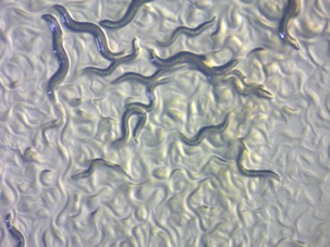
Figure 1: C. elegans in Agar under Microscope Magnification
Source: Wikimedia Commons
The paper, “Ultrasound Elicits Behavioral Responses through Mechanical Effects on Neurons and Ion Channels in a Simple Nervous System” (Jan Kubanek) discusses the mechanism by which ultrasound can stimulate a response from certain neurons in the brain of the roundworm, C. elegans (Kubanek, 2020). Kubanek used genetic knockouts to determine the neurons, ion channels, and proteins involved in ultrasound sensation. This research is highly relevant because this technique could be used to stimulate and non-invasively image deep brain structures in humans. My article begins with a background on wave mechanics and experimental set up before highlighting the key takeaways from the paper. It will conclude with future directions of the field and possible applications of this technology.
The lab used C. elegans as a model for several reasons. First, the worms are extremely sensitive to thermal and mechanical stimuli, which allowed the researchers to determine the exact mechanism of ultrasound stimulation. Second, behaviors can be tracked with cameras and software such as the Parallel Worm Tracker system used in this study. This allowed the researchers to quantitatively define behavioral responses for analysis. Third, the C. elegans nervous system has been well defined previously, allowing for the experimental data to be better understood in the context of prior research (Worm Book, 2020). Lastly, many mutants are available to study to determine how ultrasound response depends on particular proteins (Kubanek, 2020).
Worms were placed on a nematode growth agar above a piezoelectric transducer. The transducer utilizes the piezoelectric effect, the idea that electricity and force can be interconverted. Other equipment included a function generator, Arduino uno, amplifier, and a preamplifier to shape the ultrasound waves and determine which variables (duty cycle, amplitude, frequency, etc.) can be sensed and affect behavior. I explain each of these variables below. Fiber Optics were used to measure temperature generated by the ultrasound and rule out a thermal mechanism for behavioral response (Kubanek, 2020).
The genius of this paper is that all the genetic experiments mirrored each other. A mutant was chosen and then its behavior was studied. The researchers used the Parallel Worm Tracker Software to define what they called the “reversal behavior”. During ultrasound bursts, they compared the velocity vectors of the worms. The researchers defined a “reversal behavior” as a significant change in the velocity vector. After 10 trials they determined if the differences in vectors were significant. Using this method, the lab counted the number of reversal behaviors displayed by the worms. This method was carried out at different pressure to voltage ratios to determine if these variables affected stimulation (Kubanek, 2020).
The researchers shaped waves, as explained above, to determine how several variables affected stimulation. A wave’s duty cycle is the ratio of “on” versus “off”. This is analogous to automatic lights that turn off once someone leaves the room. A high duty cycle would keep the lights on long after you left the room, while a low duty cycle would quickly turn the lights on and off as you entered and exited. The pulse duration measures the amount of time the worms were subject to ultrasound bursts. The pressure variable measured the amplitude, or strength, of the ultrasound waves. The pulse repetition frequency measured the number of “reversal behaviors” at a specific frequency over a specific time in order to compare a consistent, quantitative measurement throughout the study (How Does Modulation Work?, Tait). MATLAB’s k-wave package, a computer software, was used to shape and simulate the ultrasound waves while controlling the several variables. These variations themselves were modulated into an input signal, a wave with the specific variable changes, which was them propagated and amplified through a carrier wave. This allowed the researchers to analyze how different aspects of wave forms affect behavior in worms (Kubanek, 2020).
Two preliminary experiments gave the researchers insight into the topic at hand. In the first preliminary experiment, the researchers compared a sham signal (0.0 MPa) with a bona fide signal (1.0 MPa). They tracked movement in the presence and absence of ultrasound pulses. It was found that ultrasound did in fact elicit a reversal behavior, a significant change in the velocity vector, in the worms as the lab hypothesized (Kubanek, 2020).
In the second preliminary experiment, the researchers studied the effect of ultrasound bursts for various durations of time. It was found that ultrasound does indeed induce a behavioral effect through stimulation and that for longer durations of ultrasound exposure, the behavior was stronger. The researchers found that 200 msec was the optimal duration and kept the duration constant for the remaining experiments (Kubanek, 2020).
Subsequently, the authors moved on to the major experimental objective – determining the molecular mechanism underlying ultrasound sensation in C. elegans. The authors predicted that the worm would be responsive to thermal, mechanical, or electrical effects. Thermal effects could act through temperature-dependent channels. Mechanical effects could impose force through oscillations or cavitation. Membrane oscillation could cause mechanical channels to open. Cavitation, a phenomenon in which gas packets in liquid collapse, was controlled and eliminated using low frequency waves. Another possibility included radiation force, the pressure of acustic intensity, that could possibly push open ion channels. The final proposed mechanism included electrical capacitance: membrane interactions could change the electrical properties of voltage or capacitance which could open ion channels (Kubanek, 2020).
First, the thermal mechanism was ruled out. Guanylate cyclase (GCY) mutants affect AFD neurons (Amphid neurons with finger-like ciliated endings) that sense temperature (How Does Modulation Work?, Tait). The mutants interrupt the G-protein signal cascade by preventing guanylate cyclase from converting GTP into cGMP. The researchers determined that thermosensation is not used to sense ultrasound; the thermal mutants were no different from the wild type worms so the ultrasound mechanism cannot act though thermal sensors (Kubanek, 2020).
They then turned to the other proposed mechanisms as listed in the figure above. In these next trials, a Mec-3 mutant was studied. This mutant lacks not only the touch receptor neurons in C. elegans but also, the PVD and FLP neurons. Unlike the thermal mutants, these worms were insensitive to ultrasound (Kubanek, 2020). This finding supports the idea that mechano-sensation of ultrasound is elicited by mec proteins in either the touch receptor neurons, PVD neurons, or FLP neurons (Kubanek, 2020).
To determine which neurons are utilized in the mechanism, another mec mutant was involved.
The Mec-4 mutant lacks functional touch receptor neurons but have functional PDV and FLP neurons. These worms were also not responsive to ultrasound. This shows that the mechano-sensing mechanism acts through these touch receptor neurons, a key finding of the paper (Kubanek, 2020).
The authors decided to look at another neuronal protein, TRP, known to act as a mechano-sensor as well. The TRP proteins are encoded in CEP neurons, also known as cephalic neurons (How Does Modulation Work?, Tait). The mutants were similar to wildtype worms in response to ultrasound, showing that trp-4 and CEP do not play a role in the mechanism. However, the mutants showed unpredicted side effects such as stunted growth and slower movements. The researchers decided to look at other trp mutants instead (Kubanek, 2020). The two other mutants showed no side effects and were also no different from the wild type. Therefore, the researchers were able to conclude that TRP proteins and CEP neurons do not play a role in the ultrasound sensing mechanism.
In summary, the researchers tested three possible mechanisms: thermal, mechanical, and electrical. The GCY mutants allowed the researchers to rule out temperature. Similarly, because Mec-4 is voltage-independent they were able to rule out electrical membrane interactions. In conclusion, the paper found that acoustic radiation mechanically opens the Mec-4 subunits that activate the touch receptor neurons when elicited with ultrasound. Mec-4 needs 50-100 nN to open and the MATLAB simulation predicted the neurons were getting around 900 nM of force.
Future experiments could include studying similar mechanisms in Piezo channels in mammals, K2P channels in Xenopus (African clawed frog), and NOMPC channels in the fruit fly (Kubanek, 2020). These structures display similarities to TRN in C. elegans and may provide a means for cellular stimulation. The long term goal of this technology is to allow for noninvasive cell stimulation of neurons and perhaps glia. For example, methods utilizing Sonogenetics could be employed to replace Parkinson’s disease treatments such as deep brain stimulation. Instead of surgically implanting a stimulator, an external transducer and amplifier could create and shape sound waves which could activate genetically edited neurons in the basal ganglia (Sonogenetics, Salk Institute). As Optogenetics users light to turn off and on genes, Sonogenetics, using ultrasound, may one day be able to activate genes with sound (Kubanek, 2020). Sonogenetics creates a potential and an unmatched efficiency for future experiments. Kubanek’s research provides the first step toward such a technique by determining that a mechanical, not a thermal, mechanism is responsible for ultrasound stimulation of neurons in C. Elegans.
References
How does modulation work? Tait Radio Academy. Retrieved November 10, 2020, from https://www.taitradioacademy.com/how-does-modulation-work-1-1/
Kubanek, J., Shukla, P., Das, A., Baccus, S. A., & Goodman, M. B. (2018). Ultrasound Elicits Behavioral Responses through Mechanical Effects on Neurons and Ion Channels in a Simple Nervous System. The Journal of Neuroscience, 38(12), 3081–3091. https://doi.org/10.1523/JNEUROSCI.1458-17.2018
WormBook. Retrieved November 10, 2020, from http://www.wormbook.org/
Sonogenetics. Salk Institute for Biological Studies. Retrieved December 20, 2020, from https://sonogenetics.salk.edu/
Related Posts
Intergenerational Trauma: How a history of pain brings biological costs
Source: Nicolas Raymond, CC BY 2.0 A few years ago,...
Read MoreEpigenetics: How DNA Gives Birth to Life
Figure 1: Researchers at the Planck Institute for Molecular Genetics...
Read MoreAre We Driving Our Dogs Sick Through Our Urban Lifestyles?
Figure 1: Allergic disorders in humans and dogs are associated...
Read MoreMichael Del Sesto


