
Abstract
Across the United States, Black and Hispanic patients, uninsured individuals, lower-income populations, and people living in rural areas are disproportionately affected by delays in thyroid cancer diagnosis. With early detection, five-year survival rates exceed 99% but fall to 50-71% once metastasized. Thyroid nodules occur in up to 68% of adults, with malignancy rates of 1-5% in the general population and 7-15% in clinically selected populations. Diagnosis relies on time-intensive ultrasound interpretation by radiologists using the American College of Radiology Thyroid Imaging Reporting and Data System (ACR TI-RADS), creating challenges for early diagnosis in medically underserved populations.
Research shows that AI can expedite diagnosis, but convolutional neural network (CNN)-based thyroid cancer detection is restricted by limited high-quality datasets to train CNNs. Today, traditional augmentation and Generative Adversarial Network (GAN)-generated synthetic data are used to improve training in research settings.
This study presents a novel Saliency-Guided Augmented Data Framework (SGAD-F) using model-derived saliency maps to guide augmented image generation for CNN training which preserves diagnostically meaningful regions throughout the augmentation process. CNNs trained while using SGAD-F outperformed ACR TI-RADS across all recorded performance metrics and exceeded other CNNs trained using existing data generation methods in the most clinically important metrics, including sensitivity, NPV, and LR-. These SGAD-F-guided CNNs introduce the possibility of efficient thyroid cancer diagnosis in underserved populations. Further, SGAD-F could aid in the diagnosis of rarer conditions by expanding limited datasets with high-quality data for CNN training.
CNNs trained with SGAD-F-guided data may allow for earlier thyroid cancer diagnosis in populations who currently face disproportionate barriers to timely diagnosis.
Keywords: Thyroid Cancer, Diagnostic Equity, Medical Imaging, Ultrasound Images, Data Augmentation, Limited Datasets, Convolutional Neural Networks, Saliency Maps, Grad-CAM, Explainable Artificial Intelligence, Image Classification, Medical Artificial Intelligence
Introduction
Thyroid cancer is highly treatable when detected early, making timely identification of suspicious thyroid nodules clinically important. Thyroid cancer is a disease in which abnormal cells in the thyroid gland grow uncontrollably and form a malignant tumor. Thyroid nodules are common, with ultrasound detecting nodules in as many as 68% of adults (Guth et al., 2009). However, the vast majority are benign, with an estimated malignancy rate of 1-5% among nodules identified in the general population, compared with 7-15% among nodules referred for specialized clinical evaluation (Durante et al., 2023; Grussendorf et al., 2022). For papillary, follicular, and medullary thyroid cancers, five-year relative survival exceeds 99% when the cancer is localized, but falls to approximately 71%, 62%, and 50%, respectively, after metastasis (American Cancer Society survival data, 2025). When a thyroid nodule is identified, ultrasound is used to determine whether additional evaluation is necessary. The American College of Radiology Thyroid Imaging Reporting and Data System (ACR TI-RADS) requires assessment of five sonographic characteristics (composition, echogenicity, shape, margin, and echogenic foci) to assign a risk category and determine whether fine-needle aspiration biopsy or imaging follow-up is recommended (Tessler et al., 2017).
However, access to timely thyroid cancer evaluation is not equal across the United States. A population-based analysis found that Black and Hispanic patients were more likely than non-Hispanic White patients to present with metastatic thyroid cancer, while patients who were poor, uninsured, or covered by Medicaid also had higher odds of metastatic disease than privately insured patients (Harari et al., 2014). Hispanic patients have also been shown to experience significantly longer times to initial thyroid cancer treatment and surgery than non-Hispanic patients (Kowkabany et al., 2023). Lower socioeconomic status has independently been associated with more advanced thyroid cancer at the time of detection (Almubarak et al., 2021). Geographic access appears to present an additional challenge. Rural patients with thyroid cancer have been found to present with later-stage disease and experience greater disease-specific mortality (Huston-Paterson et al., 2024). Collectively, these studies suggest that racial, socioeconomic, insurance, and geographic disparities can affect when thyroid cancer is detected.
Disparities in timely diagnosis are occurring while medicine faces a radiologist shortage. These disparities in access to timely diagnosis may be further exacerbated by growing pressures on the healthcare workforce, particularly the increasing demand for radiological services. The number of medical images in the U.S. is expected to rise by about 17-27% by 2055, while the number of radiologists may not grow fast enough to keep up with demand (Christensen et al., imaging utilization, 2025; Christensen et al., radiologist supply, 2025). This creates a significant challenge for resource-constrained healthcare settings that often serve underserved populations.
Artificial intelligence (AI) offers one potential method for assisting a shrinking radiologist supply with a growing diagnostic workload. Convolutional neural networks (CNNs) can learn patterns associated with benign and malignant thyroid nodules directly from ultrasound images, and recent research indicates that AI can improve thyroid ultrasound interpretation when used as a clinical decision-support tool. In a 2024 study, AI assistance significantly improved the diagnostic performance of junior radiologists evaluating thyroid nodules in real clinical practice (Xu et al., 2024). Commercial AI systems for thyroid ultrasound have also reached clinical evaluation, including FDA-cleared tools designed to classify thyroid nodules for malignancy (Ashton et al., 2024).
A significant barrier to developing reliable thyroid ultrasound diagnostic CNNs is the availability of sufficiently large, representative, and accurately labeled training datasets. In a conventional CNN development process, a model is trained on labeled thyroid ultrasound images and evaluated using separate validation and test data. Saliency maps are sometimes generated after training to help explain where a model focused on within an ultrasound image to make its prediction. However, collecting and labeling large medical imaging datasets is expensive and time-consuming, and privacy restrictions further limit data availability. The relatively low prevalence of malignancy among thyroid nodules can also create skewed representational imbalance in naturally collected datasets. AI studies in thyroid ultrasound classification therefore rely on methods for artificially expanding the available training data.
In CNN-based thyroid imaging research today, there are two main methods used to generate additional data for training thyroid ultrasound classifying CNNs: Traditional Augmentation and Generative Adversarial Network (GAN)-generated synthetic data. Traditional data augmentation increases datasets by applying transformations such as rotation, flipping, scaling, contrast adjustment, brightness modification, or noise to existing images. A recent analysis of ultrasound augmentation found that the transformations used are often not tailored to the specific diagnostic task which can lead to lower performance in the CNNs trained with augmented data (Tupper and Gagné, 2025). GANs are neural network frameworks that generate completely new synthetic images after training on real images. However, these frameworks require substantial training data and may introduce unrealistic structures into the images they generate (Shi et al., 2024). Under both data generation methods, however, neither traditional image augmentation nor GAN-generated synthetic data guarantees that every additional training image preserves the features relevant to the original diagnosis. An augmented or GAN-generated image intended to increase data diversity could instead create distorted and unrealistic versions of important nodule characteristics.
This research paper introduces the novel Saliency-Guided Augmented Data Framework (SGAD-F), which changes the role of saliency maps from primarily a post-training explanation tool to a pre-training device that allows for the guarantee of augmentations that retain key diagnostic features. SGAD-F first generates a saliency map from an original thyroid ultrasound image using a previously trained CNN. This initial CNN does not need to be highly accurate, provided that its classification accuracy exceeds chance according to the biased binomial test (Noirhomme et al., 2014). This is particularly relevant for expanding work on small datasets. The individual augmentations are applied to the original image, and saliency maps are generated for each of the augmented images. The augmentations are then evaluated by comparing the saliency maps of the augmented images to the saliency maps of the original images. SGAD-F compares the corresponding saliency maps through Grad-CAM, a Python library, allowing the framework to measure whether the model continues to attend to the same diagnostically relevant regions after augmentation. To quantify this preservation of diagnostic attention, SGAD-F introduces objective evaluation metrics based on saliency similarity. Spatial overlap between diagnostic regions is measured using Intersection over Union (IoU), while global similarity of attention distribution is measured using Class Activation Map (CAM) Correlation. Together, these metrics assess whether an augmentation maintains the diagnostic characteristics of the original image. High agreement between saliency maps of an augmented and an original image suggests that the transformation preserves clinically meaningful information, whereas low agreement indicates that the augmentation may alter or obscure features important for diagnosis. Augmentations that resulted in images with high IoU and CAM Correlation scores are selected as SGAD-F augmentations for training CNNs.
This study evaluates SGAD-F as a method for generating a more diagnostically consistent augmented training set for thyroid ultrasound classification. CNNs trained with SGAD-F-guided data were compared with CNNs trained using existing data-generation approaches and with ACR TI-RADS using clinically relevant performance measures including accuracy, sensitivity, negative predictive value, and negative likelihood ratio.
Results
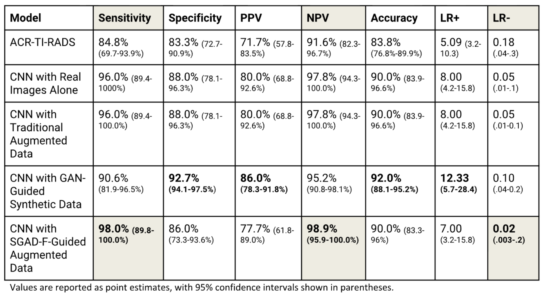
The SGAD-F model achieved 98.0% sensitivity, 86.0% specificity, 77.7% positive predictive value, 98.9% negative predictive value, and 90.0% accuracy, with a positive likelihood ratio of 7.00 and a negative likelihood ratio of 0.02.
Compared with ACR TI-RADS and CNNs trained using real images alone, traditional augmentation, or GAN-generated data, SGAD-F achieved the highest sensitivity and negative predictive value and the lowest negative likelihood ratio, indicating the strongest performance in minimizing missed thyroid cancers. However, other models performed better on specificity, positive predictive value, accuracy, and positive likelihood ratio. Figure 1 shows all of the recorded performance metrics for all five approaches.
Materials and Methods
Overview
This study evaluated five approaches to thyroid cancer detection using ultrasound imaging: ACR TI-RADS, a CNN trained on real ultrasound images alone, a CNN trained on real images supplemented with traditional augmentation, a CNN trained on real images supplemented with SGAD-F guided augmentation, and a CNN trained on real images supplemented with GAN-generated synthetic data.
Two of the five approaches were based on published studies. The ACR TI-RADS results were based on a 2021 study and the results from a CNN trained on real images supplemented with GAN-generated synthetic data were based on a 2020 study (Baz et al., 2021; Shi et al., 2020). The other three CNNs were trained on the same dataset using different augmentation approaches: no augmentation, traditional augmentation, and SGAD-F-guided augmentation. The diagnostic performance for all five approaches was compared using sensitivity, specificity, positive predictive value (PPV), negative predictive value (NPV), accuracy, and likelihood ratios. All performance metrics from this study were standardized to a disease prevalence (P*) of 33.3%, matching the prevalence used in the ACR TI-RADS performance metrics reported by Baz et al. in 2021. Prevalence standardization allows for fair comparison across studies with different proportions of benign versus malignant nodules being assessed. To standardize, Bayes’ theorem was used to recompute PPV, NPV, and accuracy using the study’s sensitivity and specificity (Shi et al., 2020).
Confidence intervals (CIs) for each diagnostic performance metric were calculated to better represent uncertainty in the reported performance metrics. 95% CIs were estimated using bootstrapping, with ChatGPT used to perform repeated resampling of the test dataset with replacement (20,000 iterations) and to recalculate sensitivity, specificity, PPV, NPV, accuracy, and likelihood ratios. The 2.5th and 97.5th percentiles of the resulting distributions defined the 95% confidence intervals, providing a more reliable measure of model performance than single point values.
ACR TI-RADS Study
The ACR TI-RADS diagnostic performance metrics were documented from a publicly available clinical study (Baz et al., 2021). The American College of Radiology Thyroid Imaging Reporting and Data System (ACR TI-RADS) is a standardized ultrasound-based framework for assessing thyroid nodules. It assigns points based on five imaging characteristics: composition, echogenicity, shape, margins, and echogenic foci. The total score classifies nodules from TR1 (benign) to TR5 (highly suspicious), guiding recommendations for biopsy or ultrasound follow-up based on risk category and nodule size (Tessler et al., 2017). When selecting the diagnostic performance metrics for the ACR TI-RADS model, the metrics based on the TR4+ threshold were used. TR4+ includes nodules classified as moderately suspicious (TR4) or highly suspicious (TR5) based on their ultrasound characteristics. The TR4+ threshold is widely used when clinicians prioritize not missing cancers. Multiple studies show that using TR4+ rather than only TR5 catches more malignant nodules (higher sensitivity), while still having a strong specificity, avoiding false positives. Using TR3 only would bring in too many benign nodules (too many false positives).
GAN-Generated Data Study
Diagnostic performance metrics for the CNN trained with GAN-generated data were obtained from the published study by Shi et al. in 2020, which evaluated a CNN using GAN-generated synthetic thyroid ultrasound images.
CNN Training
The three other approaches compared in this study consist of CNNs that were trained on three dataset variations: no augmentation, traditional augmentation, and SGAD-F-guided augmentation. All CNNs were trained in the Python programming language (Supplementary Method S1).
Dataset
Two publicly available thyroid ultrasound image datasets made up the images that were used to train and test CNNs: Algerian Ultrasound Images Thyroid Dataset (AUITD) and the Thyroid Cancer Classification Ultrasound Dataset on Kaggle (Azouzmaroua, n.d.; Diveshzz, n.d.). The images in these datasets have no annotations on them, as markings can easily mislead the model during training. The images used were split into training, validation, and testing sets to prevent bias. There were 300 base images per class with 200 for training, 50 for validation, and 50 for testing. The images were of papillary thyroid carcinoma, which accounts for about 80% of thyroid cancers according to the American Cancer Society and is the most represented subtype in publicly available ultrasound datasets. The ultrasound features used to inform biopsy decisions are the same for all cancer subtypes. Since these diagnostic features do not change based on subtype, a model trained on papillary cancer remains relevant and reliable for detecting thyroid cancer risk in general.
Hyperparameter Grid Search
Hyperparameter grid search was used to evaluate different image resolutions, batch sizes, and learning rates. The tested values are described in Supplementary Method S2.
Preprocessing
Before the training cycle, images were cropped, resized based on the model’s hyperparameters, and the values were normalized. Normalization involves scaling each image’s pixel values to a common small range (0-1 by dividing by 255) so inputs are consistent and the network learns faster and with better accuracy.
Traditional Augmentation
In this study, the traditional augmented data was generated in the following way:
- A set of standard transformations were selected
- For every image in the training set, one transformation from the set was selected at random and applied
- Step 2 was repeated, tripling the amount of images in the training set
The set of standard transformations used on the thyroid ultrasound images were zoom, rotation, vertical flipping, horizontal flipping, brightness, elastic transformation, and gaussian noise, all of which were among the 12 most commonly used augmentations observed in Tupper and Gagné’s 2025 study.
SGAD-F
a. Prerequisites
Before implementing SGAD-F, a model that was already trained on the same class and dataset to be augmented with SGAD-F was necessary. Extremely high performance was not required for this initial model, provided that its classification accuracy exceeded chance according to the biased binomial test described by Noirhomme et al. The highest performing CNN trained with no augmentation, which was also one of the five approaches being compared in this study, served as the initial model when implementing SGAD-F.
b. Saliency Map Generation
A set of 10 images in the initial CNN’s testing set was selected randomly. These images were diagnosed by the initial CNN. After diagnosis, a saliency map was generated using Grad-CAM to identify the regions of an image the CNN focused on when making its prediction. In a saliency map, each pixel is assigned a value representing its importance to the model’s decision, with higher importance shown as brighter pixels. Individual image augmentations were then applied to the original image, those images were diagnosed, and then new saliency maps were generated for each augmented version. 11 transformations were applied at three different intensities for 33 total augmentation instances. These 33 instances were applied to the 10 selected thyroid ultrasound images, and saliency maps were then generated for the 10 original images and their 330 augmented versions using the CNN mentioned in the Prerequisites section.
c. Augmentation Evaluation
Augmentations were then evaluated by comparing the corresponding saliency maps through Grad-CAM, allowing the framework to measure whether the model continued to attend to the same diagnostically relevant regions after augmentation. To quantify this preservation of diagnostic attention, SGAD-F introduced objective evaluation metrics based on saliency similarity. Spatial overlap between diagnostic regions was measured using Intersection over Union (IoU), while global similarity of attention distribution was measured using Class Activation Map (CAM) Correlation. High agreement between saliency maps of an augmented and an original image suggested that the transformation preserved clinically meaningful information, whereas low agreement indicated that the augmentation may have altered or obscured features important for diagnosis. Augmentations that resulted in images with high IoU and CAM Correlation scores across the majority of the 10 images were selected as SGAD-F augmentations for training CNNs.
This step was performed on the 340 total thyroid ultrasound images, with each of the 330 augmented images being individually compared to their original counterparts. Out of the 33 individual transformations, 13 of them were selected as SGAD-F augmentations for thyroid nodule classification.
Performance Metrics
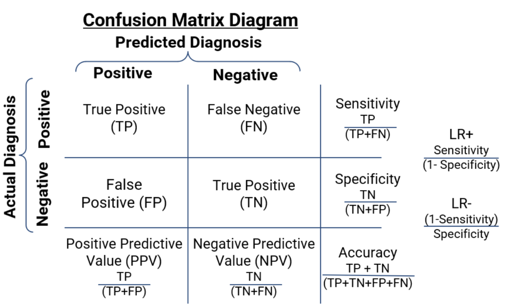
The three most clinically important performance metrics are sensitivity, negative predictive value (NPV), and negative likelihood ratio (LR-). Higher sensitivity reduces missed cancers, higher NPV increases confidence in negative results, and a lower LR- sharply decreases post-test cancer probability after a negative prediction. These metrics are the most clinically important because a false negative is much more detrimental to a patient than a false positive, and the three metrics focus on reducing false negatives and increasing true positives. In addition to these three metrics, accuracy, specificity, positive predictive value (PPV), and positive likelihood ratio (LR+) were recorded. These values were derived from each approach’s confusion matrix, as shown in Figure 2, which displays the number of true positives, true negatives, false positives, and false negatives produced by each approach which is an indicator of the performance.
Discussion
As seen in Figure 1, the CNN trained using the novel SGAD Framework outperformed all other evaluated approaches in sensitivity, negative predictive value (NPV), and negative likelihood ratio (LR-), three of the most clinically important measures for ruling out thyroid cancer (highlighted). Higher sensitivity reduces the risk of missed cancers, while higher NPV and a lower LR- increase confidence that a negative result truly indicates a low probability of malignancy.
One possible reason for the improved performance is that SGAD-F selected augmentation transformations based on whether they preserved the image regions that the CNN considered diagnostically important. Traditional augmentation applies predefined transformations without confirming that important thyroid nodule features remain intact. In contrast, SGAD-F used Grad-CAM-based saliency comparisons to identify transformations that maintained similar regions of model attention between the original and augmented images. Training on these more diagnostically consistent augmented images may have allowed the CNN to gain the benefits of additional training data without introducing high levels of irrelevant or distorted information.
This feature may also have important implications for clinical adoption. Although AI systems for thyroid ultrasound are increasingly being developed and several platforms have received FDA clearance, widespread clinical use remains limited. A major concern with CNNs is their “black-box” nature: clinicians may receive a prediction without understanding which image features contributed to it. Lack of explainability has been repeatedly identified as a barrier to clinical trust and adoption. SGAD-F does not make the CNN fully explainable, but it introduces greater transparency into the training process by demonstrating that the augmented images retained diagnostically important regions identified by the model. This could make the augmentation process more clinically understandable and may support greater confidence in models trained with SGAD-F.
SGAD-F may also be useful when training data are limited. CNNs require large and diverse datasets, which can be difficult to obtain for uncommon cancers or rare disease subtypes. A framework that identifies clinically appropriate transformations could expand smaller datasets without requiring completely new patient images. In particular, this may be valuable for rare cancers, where collecting enough real cases to train high-performing CNNs can be difficult.
The potential clinical impact extends beyond diagnostic performance. AI-assisted thyroid ultrasound could help radiologists interpret growing imaging volumes more efficiently and potentially reduce workload. Previous work has suggested that AI could improve both the accuracy and efficiency of thyroid ultrasound interpretation. If SGAD-F-guided CNNs maintain strong performance across larger and more diverse clinical datasets, they could eventually serve as decision-support tools in settings with limited radiology resources. This could have important applications for underserved populations, where shortages of specialized healthcare resources may contribute to delays in diagnosis.
Several limitations should be considered. SGAD-F was evaluated using a relatively limited number and range of datasets, and testing across larger, more diverse, and external datasets would be necessary to determine whether its performance generalizes across patient populations, ultrasound machines, institutions, and thyroid cancer subtypes. The SGAD-F-guided CNN also did not outperform the GAN-based approach on every diagnostic metric. GAN-generated data therefore remain an important alternative. However, GANs themselves require real training data to learn the underlying image distribution, can require substantial annotated datasets and computational resources, and may introduce synthetic artifacts or clinically unrealistic features. This may reduce their practicality in exactly the data-limited settings where augmentation is most needed.
Future studies should evaluate SGAD-F across larger and more diverse thyroid ultrasound datasets and compare it prospectively with traditional augmentation, GAN-generated data, and standard clinical approaches. External validation and testing in real clinical workflows will be particularly important to determine whether SGAD-F’s improvements in sensitivity, NPV, and LR- translate into fewer missed cancers, greater clinician confidence, and improved diagnostic efficiency.
Conclusion
This study found that the CNN trained using the novel SGAD-F framework achieved the strongest performance in sensitivity, negative predictive value, and negative likelihood ratio, the three metrics most important for reducing missed cancers and increasing confidence in negative results. By selecting augmentation techniques that preserved diagnostically important image regions, SGAD-F provided a more targeted alternative to standard augmentation while also adding greater transparency to the model-training process.
By combining augmentation with saliency-based quality control, SGAD-F has the potential to improve the reliability of CNN training for more efficient thyroid cancer diagnosis, potentially supporting earlier identification of suspicious nodules in populations that currently experience disproportionate barriers to timely care. The same framework may also have broader applications in rare cancers and other medical imaging tasks in which limited labeled data constrain the development of reliable diagnostic CNNs.
Acknowledgments
I would like to express my gratitude for my mentor, Barbara Morrow, for her guidance, encouragement, and advice throughout the process of my research. Her mentorship has been key to supporting the direction of my research, and I am truly thankful for her guidance.
Supplementary Information
Supplementary Method S1. CNN Training
All CNN models were trained using the EfficientNet-B2 architecture, 600 thyroid ultrasound images, hyperparameter grid searching, and standard image preprocessing for CNNs. EfficientNet-B2 was selected for its ability to achieve strong image classification performance while maintaining computational efficiency.
Supplementary Method S2. Hyperparameter Grid Search
The specific hyperparameters tested in this study were as follows: 160 by 160, 220 by 200, or 300 by 300 pixels image size, 4, 8, or 12 images per batch while training, and 1e-4, 5e-4, 1e-3, 1.5e-3, or 2e-2 learning rate.
References
Almubarak, A. A., Albkiry, Y. A., Alsalem, A. A., & Elkrim Saad, M. A. (2021). The association of low socioeconomic status with advanced stage thyroid cancer. Journal of Taibah University Medical Sciences, 16(4), 482-490. https://doi.org/10.1016/j.jtumed.2021.04.006
American Cancer Society. (n.d.). Thyroid cancer survival rates. https://www.cancer.org/cancer/types/thyroid-cancer/detection-diagnosis-staging/survival-rates.html
Ashton, J., Morrison, S., Erkanli, A., & Wildman-Tobriner, B. (2024). Assessment of the Diagnostic Performance of a Commercially Available Artificial Intelligence Algorithm for Risk Stratification of Thyroid Nodules on Ultrasound. Thyroid: Official Journal of the American Thyroid Association, 34(11), 1379-1388. https://doi.org/10.1089/thy.2024.0410
American Cancer Society. (2025, June 30). Thyroid cancer survival rates, by type and stage. https://www.cancer.org/cancer/types/thyroid-cancer/detection-diagnosis-staging/survival-rates.html
Azouzmaroua. (n.d.). Algerian Ultrasound Images Thyroid Dataset: AUITD [Dataset]. Kaggle. https://www.kaggle.com/datasets/azouzmaroua/algeria-ultrasound-images-thyroid-dataset-auitd
Baz, A. A., Mohamed, A. H. I., El-Esawy, Y. F. G., & El-kaffas, K. H. (2021). Conventional ultrasound, color Doppler, TI-RADS, and shear wave elastography for thyroid nodule differentiation: A study of efficacy compared with the histopathology results. Egyptian Journal of Radiology and Nuclear Medicine, 52, Article 103. https://doi.org/10.1186/s43055-021-00474-5
Christensen, E. W., Drake, A. R., Parikh, J. R., Rubin, E. M., & Rula, E. Y. (2025). Projected US Imaging Utilization, 2025 to 2055. Journal of the American College of Radiology: JACR, 22(2), 151-158. https://doi.org/10.1016/j.jacr.2024.10.017
Christensen, E. W., Parikh, J. R., Drake, A. R., Rubin, E. M., & Rula, E. Y. (2025). Projected US Radiologist Supply, 2025 to 2055. Journal of the American College of Radiology: JACR, 22(2), 161-169. https://doi.org/10.1016/j.jacr.2024.10.019
Durante, C., Hegedüs, L., Czarniecka, A., Paschke, R., Russ, G., Schmitt, F., Soares, P., & Solymosi, T. (2023). 2023 European Thyroid Association clinical practice guidelines for thyroid nodule management. European Thyroid Journal, 12(5), e230067. https://doi.org/10.1530/ETJ-23-0067
Diveshzz. (n.d.). Thyroid cancer classification ultrasound dataset [Dataset]. Kaggle. https://www.kaggle.com/datasets/diveshzz/thyroid-cancer-classification-ultrasound-dataset
Grussendorf, M., Ruschenburg, I., & Brabant, G. (2022). Malignancy rates in thyroid nodules: a long-term cohort study of 17,592 patients. European Thyroid Journal, 11(4), e220027. https://doi.org/10.1530/ETJ-22-0027
Guth, S., Theune, U., Aberle, J., Galach, A., & Bamberger, C. M. (2009). Very high prevalence of thyroid nodules detected by high frequency (13 MHz) ultrasound examination. European Journal of Clinical Investigation, 39(8), 699–706. https://doi.org/10.1111/j.1365-2362.2009.02162.x
Harari, A., Li, N., & Yeh, M. W. (2014). Racial and socioeconomic disparities in presentation and outcomes of well-differentiated thyroid cancer. The Journal of Clinical Endocrinology and Metabolism, 99(1), 133-141. https://doi.org/10.1210/jc.2013-2781
Huh, S., Lee, H. S., Yoon, J., Kim, E.-K., Moon, H. J., Yoon, J. H., Park, V. Y., & Kwak, J. Y. (2020). Automatic classification of thyroid nodules from ultrasound images using deep learning networks. Scientific Reports, 10, Article 5602. https://doi.org/10.1038/s41598-020-67543-z
Huston-Paterson, H. H., Mao, Y., Tseng, C. H., Kim, J., Bobanga, I., Wu, J. X., & Yeh, M. W. (2024). Rural-Urban Disparities in the Continuum of Thyroid Cancer Care: Analysis of 92,794 Cases. Thyroid: Official Journal of the American Thyroid Association, 34(5), 635-645. https://doi.org/10.1089/thy.2023.0357
Kowkabany, G., Jimenez, P. B., Sharan, S., Bansal, S., Ahmed, S. H., Raikot, S., Manaise, H. K., Popp, R., Popp, K., Sukniam, K. B., & Gabriel, E. (2023). Inequities in Time to Treat Thyroid Cancer. Anticancer Research, 43(11), 5025-5030. https://doi.org/10.21873/anticanres.16701
Megwalu, U. C., & Ma, Y. (2021). Racial/Ethnic Disparities in Use of High-Quality Hospitals Among Thyroid Cancer Patients. Cancer Investigation, 39(6-7), 482-488. https://doi.org/10.1080/07357907.2021.1938108
Noirhomme, Q., Lesenfants, D., Gomez, F., Soddu, A., Schrouff, J., Garraux, G., Luxen, A., Phillips, C., & Laureys, S. (2014). Biased binomial assessment of cross-validated estimation of classification accuracies illustrated in diagnosis predictions. NeuroImage: Clinical, 4, 687-694. https://doi.org/10.1016/j.nicl.2014.04.004
Neiman Health Policy Institute. (n.d.). New studies shed light on the future radiologist workforce shortage by projecting future radiologist supply and demand for imaging. https://www.neimanhpi.org/press-releases/new-studies-shed-light-on-the-future-radiologist-workforce-shortage-by-projecting-future-radiologist-supply-and-demand-for-imaging/
Shi, G., Wang, J., Qiang, Y., Yang, X., Zhao, J., Hao, R., Yang, W., Du, Q., & Kazihise, N. G.-F. (2020). Knowledge-guided synthetic medical image adversarial augmentation for ultrasonography thyroid nodule classification. Computer Methods and Programs in Biomedicine, 196, Article 10561. https://doi.org/10.1016/j.cmpb.2020.105611
Tareke, T. W., Leclerc, S., Vuillemin, C., Buffier, P., Crevisy, E., Nguyen, A., Monnier Meteau, M. P., Legris, P., Angiolini, S., & Lalande, A. (2024). Automatic Classification of Nodules from 2D Ultrasound Images Using Deep Learning Networks. J Imaging, 10(8), 203. https://doi.org/10.3390/jimaging10080203
Tessler, F. N., et al. (2017). ACR Thyroid Imaging, Reporting and Data System (TI-RADS): White Paper of the ACR TI-RADS Committee. Journal of the American College of Radiology: JACR, 14(5), 587-595. https://doi.org/10.1016/j.jacr.2017.01.046
Tupper, A., & Gagné, C. (2025). Revisiting data augmentation for ultrasound images (arXiv:2501.13193v1) [Preprint]. arXiv. https://arxiv.org/abs/2501.13193
Xu, D., Sui, L., Zhang, C., Xiong, J., Wang, V. Y., Zhou, Y., Zhu, X., Chen, C., Zhao, Y., Xie, Y., Kong, W., Yao, J., Xu, L., Zhai, Y., & Wang, L. (2024). The clinical value of artificial intelligence in assisting junior radiologists in thyroid ultrasound: a multicenter prospective study from real clinical practice. BMC Medicine, 22(1), 293. https://doi.org/10.1186/s12916-024-03510-z
Wu, G. G., Lv, W. Z., Yin, R., Xu, J. W., Yan, Y. J., Chen, R. X., Wang, J. Y., Zhang, B., Cui, X. W., & Dietrich, C. F. (2021). Deep Learning Based on ACR TI-RADS Can Improve the Differential Diagnosis of Thyroid Nodules. Front Oncol, 11, 575166. https://doi.org/10.3389/fonc.2021.575166
Zagzag, J., Kenigsberg, A., Patel, K. N., Heller, K. S., & Ogilvie, J. B. (2017). Thyroid cancer is more likely to be detected incidentally on imaging in private hospital patients. The Journal of Surgical Research, 215, 239-244. https://doi.org/10.1016/j.jss.2017.03.059
When the Going gets Tough, the Close start Nearing: Networks and External Stress
Figure: A Social Network (Wikimedia Commons, Author Zigomitros Athanasios –...
Read More
Risk of Death Among Recently Released Inmates
Figure 1: While healthcare inequity is certainly an issue while...
Read More
The Geography of Disease: a Bayesian Approach to Epidemiology
Figure 1: A map showing relative rates of pancreatic cancer...
Read More
A Novel Mental Health Literacy Education Program for Pre-Teens
Figure: A typical elementary school classroom in Japan (Cassidy, 2005). Source:...
Read More
Autism Spectrum Disorder and Gender: The Case for Expanding the Autistic Phenotype
This article was originally submitted to the Modern MD competition...
Read More
The H.U.N.G.E.R. Initiative (Helping Underserved Neighborhoods Generate Equitable Resources)
This publication is in proud partnership with Project UNITY’s Catalyst Academy 2023...
Read More