
September 24, 2021

How Tele-Mental Health Helps Us During the COVID-19 Pandemic
Figure 1: Telehealth consultation via smartphones can serve as a...
Read More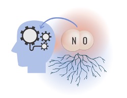
There’s NO Telling What Nitric Oxide Might Bring to Alzheimer’s Disease Research
Figure 1: As Alzheimer’s disease research begins to shift away...
Read More
Early Childhood Adversity Impacts Impulsive Decision-Making
Cover Image: An image of a girl holding a stuffed...
Read More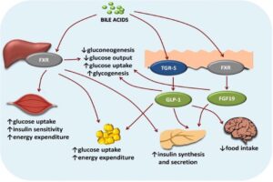
Bile Acid-Induced Satiety to Treat Obesity
Figure 1: Diagram of the effects of bile acids on...
Read More
Human Endogenous Retroviruses Might Unlock a New Field of Neurodegenerative Disease Research
Figure 1: Researchers have found viral genetic materials within the...
Read More
CellChat Enables Biologists to Understand Cellular Communication
Figure 1: CellChat, an open-source R package, provides researchers with...
Read More